
Rapid Fire Abstracts
Alexandra Jack, BSc
Student
University of Michigan
Sukran Erdem, MD
Research Assistant
UT Southwestern Medical Center

Gerald Greil, MD, PhD
Professor
UT Southwestern Medical Center

Tarique Hussain, MD, PhD
Professor
UT Southwestern
Qing Zou, PhD
CMR Physics
UT Southwestern
Qing Zou, PhD
CMR Physics
UT Southwestern

Non-contrast MR angiography allows heart and vascular imaging without contrast agents but suffers from low signal-to-noise ratio (SNR) and contrast-to-noise ratio (CNR), flow artifacts, and long acquisition times. The Relaxation-Enhanced Angiography without Contrast and Triggering (REACT) sequence improves SNR and CNR but still has challenges, including extended acquisition times and difficulty visualizing smaller vessels like pulmonary veins. Thus, further advancements in non-contrast techniques are needed for better cardiovascular imaging.
Methods:
To overcome the limitation of using ECG and respiratory-triggered REACT for imaging the pulmonary veins and/or pulmonary arteries and some other small lung vessels, we proposed the magnetization transfer contrast (MTC) REACT (MTC-REACT) in this work. With this technique, bound-pool protons are also selectively saturated, and the background signal is suppressed, improving the visibility of vascular structures, specifically the pulmonary veins and arteries [1]. To address the challenge of extended acquisition times, this study implements a deep-learning reconstruction algorithm tailored for highly undersampled data.
Twenty subjects participated in this study. For data acquisition, we used respiratory navigation and ECG triggering combined with a 3D magnetization prepared non-balanced dual echo Dixon technique, which was first proposed in [1]. The sequence starts with a set of magnetization preparations, which consist of a MT on-resonance pre-pulse [2], a four adiabatic-based T2-preparation module. Then, a non-volume-select short inversion recovery (STIR) pre-pulse with a short inversion recovery time (TI) occurs. Sequence parameters of the REACT and MTC-REACT include: TE1/TE2/TR = 1.88/4.5/6.8, flip angle = 15, acquired isotropic voxel size 1.6 mm^3, reconstruction isotropic voxel size 0.8 mm^3, undersampling factor 10. The raw k-space data were saved for the off-line compressed-sensing-based and deep-learning-based reconstruction for comparison. For deep-learning-based image reconstruction, we utilized the Adaptive-CS-Net [3], which builds upon the ISTA-Net framework [4].
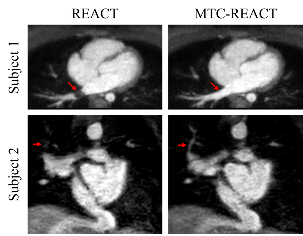
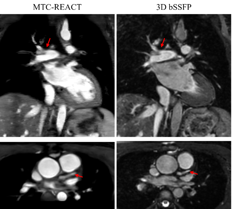
Results: Quantitative image analysis was performed using MATLAB (R2022a). To accurately assess and quantify the images, we utilized two image quality metrics including SNR and CNR. The effectiveness of the proposed MTC-REACT technique together with the deep-learning-based reconstruction framework was quantitively and qualitatively illustrated by comparing the original REACT sequence and MTC-REACT sequence. See figure 1 and figure 2. From both the quantitative and qualitative results, it can be seen that the MTC-REACT outperforms the original REACT sequence and provides better vascular visualization. In figure 3, it highlights the usage of MTC-REACT in a patient with congenital heart defect by comparing it to the contrast-enhanced 3D bSSFP sequence, which is the sequence used as clinical standard. The figure shows that the MTC-REACT and 3D bSSFP sequences provide similar information.
Conclusion:
In this work, we introduced MTC-REACT, a new non-contrast-enhanced imaging technique for whole-heart assessment, and provided preliminary insights into its application for evaluating cardiac and vascular structures. This technique showed a satisfactory visual agreement with traditional contrast-enhanced 3D bSSFP imaging. Additionally, the integration of deep-learning reconstruction facilitated rapid data acquisition.
Visual comparison between MTC-REACT sequence and the original REACT sequence. Images from two subjects comparing the original REACT (left) and MTC-REACT (right) for pulmonary veins, and pulmonary vessels were shown. The red arrows indicate clear differences between the original REACT and MTC-REACT sequences.
SNR and CNR comparison between REACT and MTC-REACT. The values shown in the table are the means of all the subjects. ** in the p-value row indicates that the corresponding p-value is less than 0.05, and * represents p-value which is larger than 0.05.
Illustration of the effectiveness of MTC-REACT by comparing to the contrast-enhanced 3D bSSFP sequence. The figure shows that the MTC-REACT and 3D bSSFP sequences provide similar information.
