Oral Abstract
Nikoo Mashayekhi, MSc
Research Assistant
McGill University, Canada
Nikoo Mashayekhi, MSc
Research Assistant
McGill University, Canada
Moezedin javad Rafiee, MD
Research associate
Research Institute of the McGill University Health Center, Canada
Mitchel Benovoy, PhD
PhD
Area 19 Medical Inc., Canada

Matthias G. Friedrich, MD
Full Professor
McGill University Health Centre
Mc Gill University, Canada
Michael Chetrit, MD
Assistant professor
McGill University Health Center, Canada
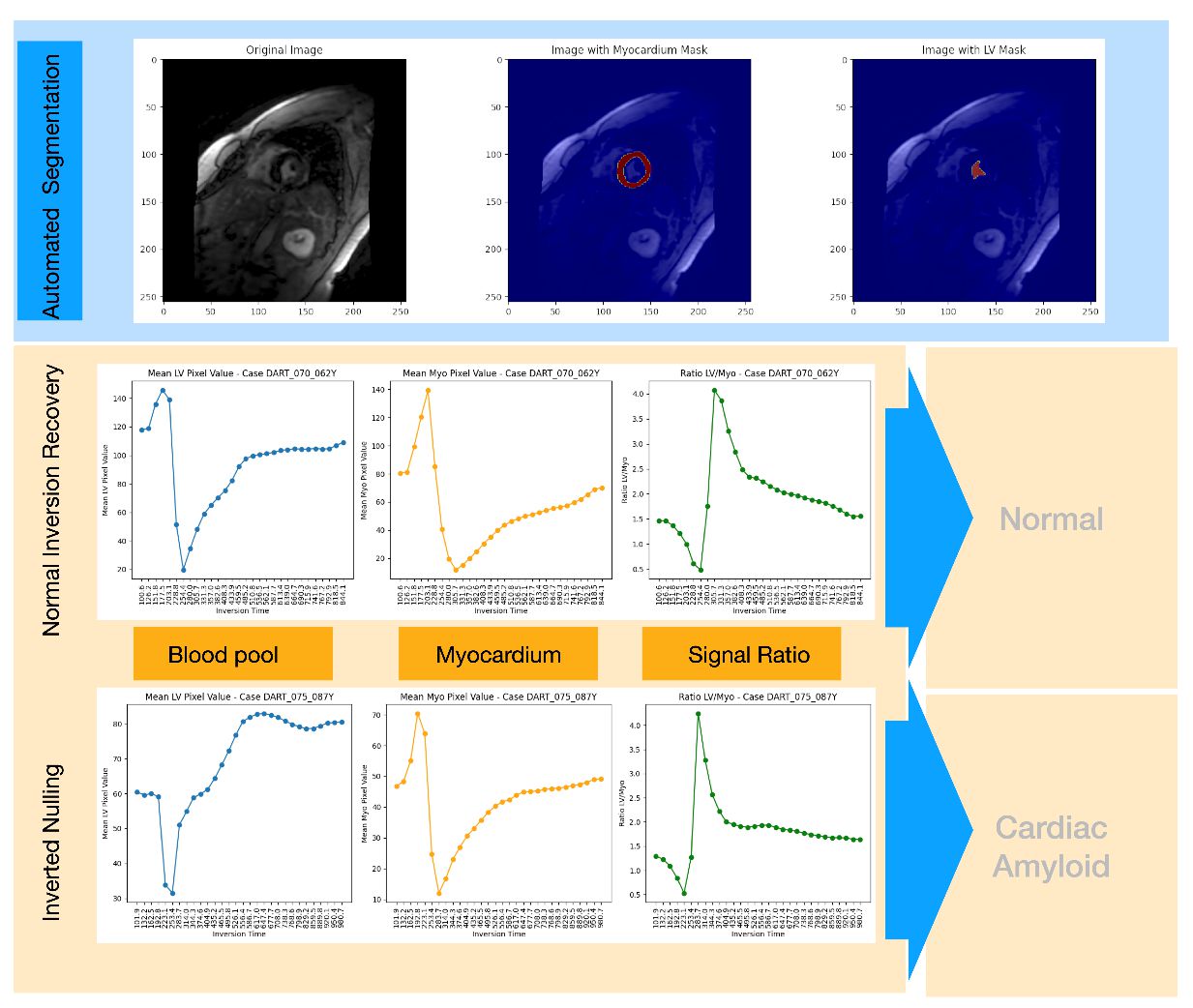
The automated TI selection method showed a mean difference of 27.12 ms earlier and a mean absolute error (MAE) of 42.17 ms compared to technologist-selected TIs, with an accuracy within 60 ms in 87.06% of cases. In 90.59% of cases, the algorithm-selected TI was at or earlier than the technologist's choice (Figure 1). In 9 out of 10 cases, expert readers preferred the -30 ms offset, noting superior contrast. Figure 2 shows examples of normal myocardium and amyloidosis. In amyloidosis, the myocardium peaks before the blood pool (reverse kinetics), contrary to the usual pattern in non-amyloidosis cases. The algorithm successfully identified this characteristic, demonstrating its ability to detect pathophysiological differences.
Conclusion: The automated segmentation and contrast optimization algorithm for delineating myocardial scarring (LGE) showed an accuracy comparable to experienced technologists, and, in some cases, provided better contrast. It also identified the altered gadolinium inflow kinetics in amyloidosis. Further validation could establish this tool as a way to improve consistency and reduce operator dependency in LGE imaging.
Figure 1 Algorithm vs Technologist-Selected TIs /Scatter plot comparing TIs selected by the algorithm and technologists. Points near the red dashed line indicate agreement, with the algorithm often selecting earlier TIs.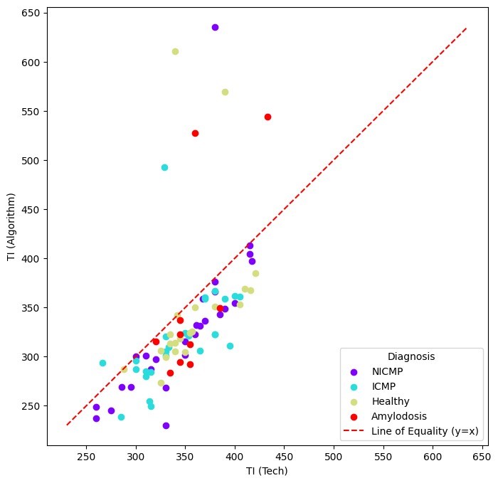
Figure 2 Automated Segmentation and Signal Analysis/ Automated segmentation of myocardium and LV blood pool, with signal intensity curves for normal myocardium and amyloidosis. Amyloidosis shows reverse kinetics, where myocardium peaks before the blood pool.