
Rapid Fire Abstracts
Moritz C. Halfmann, MD
Resident physician
University Medical Center Mainz, Germany
Moritz C. Halfmann, MD
Resident physician
University Medical Center Mainz, Germany
Lara van der Meulen, BA
PhD student
Maastricht University, Netherlands
Martijn Smulders, MD
Maastricht University Medical Center, Netherlands
Hedwig M.J.M. Nies, BA
PhD student
Maastricht University, Netherlands
Frits W. Prinzen, PhD
Professor
Maastricht University, Netherlands
Casper Mihl, MD, PhD
Assistant Professor
Maaastricht University, Netherlands
Akos Varga-Szemes, MD, PhD
Associate Professor of Radiology
Medical University of South Carolina

Robert J. Holtackers, MSc, PhD
MR Physicist
Maastricht University Medical Centre, Netherlands
Focal myocardial fibrosis is commonly qualitatively identified using late gadolinium enhancement cardiac MRI. For quantitative assessment of fibrosis, extracellular volume fraction (ECV) has emerged as a surrogate imaging biomarker. However, literature about how quantitative ECV measurements change longitudinally when acute myocardial infarction transforms into a chronic state is scarce. Therefore, the purpose of this study was to compare myocardial ECV measurements by cardiac MRI at 1 and 7 weeks following balloon catheter-induced myocardial infarction in a large animal infarct model.
Methods:
This study was approved by the Experimental Animal Committee of Maastricht University (DEC2016-002) and all animal handling complied with the Dutch Law on Animal Experimentation as well as the European Directive on the Protection of Animals used for Scientific Purposes (2010/63/EU). Directly before the start of the cardiac MRI, hematocrit levels were drawn. The MRI was performed on a 1.5T scanner (Ingenia, Philips Healthcare, Best, the Netherlands) and all animals were under general anesthesia and ventilated. Commercially available T1 maps of basal, midventricular and apical short-axis slices of the left ventricle were acquired before (5[3]3-MOLLI) and 7 minutes after (4[1]3[1]2-MOLLI) the intravenous administration of 0.2 mg/kg of gadobutrol (Gadovist, Bayer Pharmaceuticals, Berlin, Germany). Subsequently, pre- and postcontrast T1 relaxation times were quantified and ECV was calculated both globally and separately for the infarcted and remote myocardium. Results were compared using Pearson’s correlation and paired sample student’s t-tests.
Results:
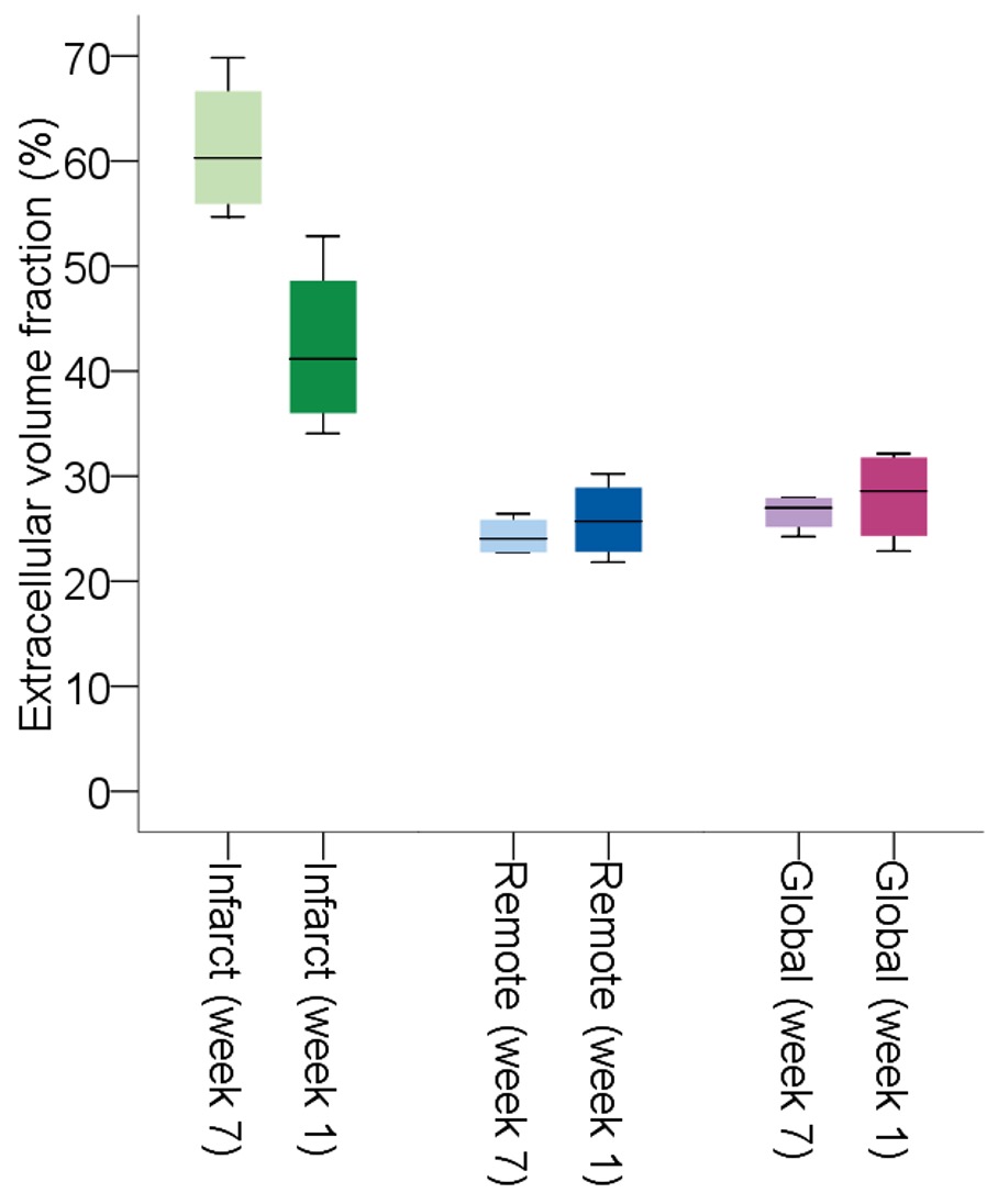
Myocardial infarction was successfully induced in all 13 Yorkshire pigs. However, 7 animals died before the week 1 MRI scan due to severe arrhythmias, 2 animals did not undergo the week 1 scan due to being unstable, and 1 animal died in between the two scans. Thus MRI at 1-week and 7-week post-infarction was successful in four and five pigs, respectively. Median time between induction of the myocardial infarction and cardiac MRI was 8 days [IQR 8 – 8] and 51 days [IQR 51 – 55]. Mean myocardial ECV was significantly higher at week 7 compared to week 1 for infarcted myocardium infarcted (61.3 ± 6.8 % vs. 42.3 ± 8.2 %, P=.011), while there was no evidence of a difference for remote (24.3 ± 1.9 % vs. 25.9 ± 3.8 %, P=.29) or global myocardium (26.5 ± 1.8 % vs. 28.0 ± 4.5 %, P=.43), respectively. All measurements showed a moderate-to-strong (r ≥ 0.62), yet statistically insignificant (P≥.16) correlation between each other.
Conclusion:
There was no difference in global myocardial extracellular volume fractions despite expected changes in areas of focal myocardial scar remodeling between 1 and 7 weeks after balloon catheter-induced myocardial infarction in a large animal model. These findings suggest looking beyond global myocardial extracellular volume fractions for a comprehensive assessment of underlying myocardial pathology.
Figure 1: Boxplots of mean global myocardial extracellular volume fraction at one and 7 weeks after catheter induced myocardial infarction for infarcted myocardium (left, green), remote myocardium (middle, blue) and global myocardium (right, purple).
