Science Sessions
Robert S. Zhang, MD
Clinical Instructor
Division of Cardiology, Weill Cornell Medicine
Robert S. Zhang, MD
Clinical Instructor
Division of Cardiology, Weill Cornell Medicine
Lily Jin, BSc
Research Coordinator
Weill Cornell Medicine
Jiahao Li, MSc
PhD Candidate
Weill Cornell Medicine
Mahniz Reza, BA
Research Assistant
Weill Cornell Medicine
Rachel Axman, MD
Resident, Internal Medicine
Weill Cornell Medicine
Pablo Villar-Calle, MD
Instructor in Medicine
Weill Cornell Medicine
Giorgia Falco, MD
MD, Fellow
Weill Cornell Medicine, Presbyterian Hospital, New York
Nil Rawal, MD
Resident
Weill Cornell Medicine
Udhay Krishnan, MD
Assistant Professor of Medicine
Weill Cornell Medicine
Nupoor Narula, MD, MSc
Assistant Professor of Medicine
Weill Cornell Medicine
Annie Tsay, MD
Fellow in Medicine
NYP-Weill Cornell Medical Center
Andre T. Cheng, MD
Clinical Fellow
Weill Cornell Medicine

Pascal Spincemaille, PhD
Associate Professor of Physics Research In Radiology
Weill Cornell Medicine

Jonathan W. Weinsaft, MD
Chief of Cardiology, Professor of Medicine
Weill Cornell Medicine

Jiwon Kim, MD
Associate Professor of Medicine
Weill Cornell Medicine
Patients with SLE show reduced stress myocardial perfusion consistent with some degree of coronary microvascular dysfunction. These findings support the use of quantitative stress myocardial perfusion CMR for assessment of coronary microvascular dysfunction in patients, potentially guiding targeted cardiovascular risk management.
Figure 1. Comparison of quantitative stress myocardial perfusion between controls and patients with SLE.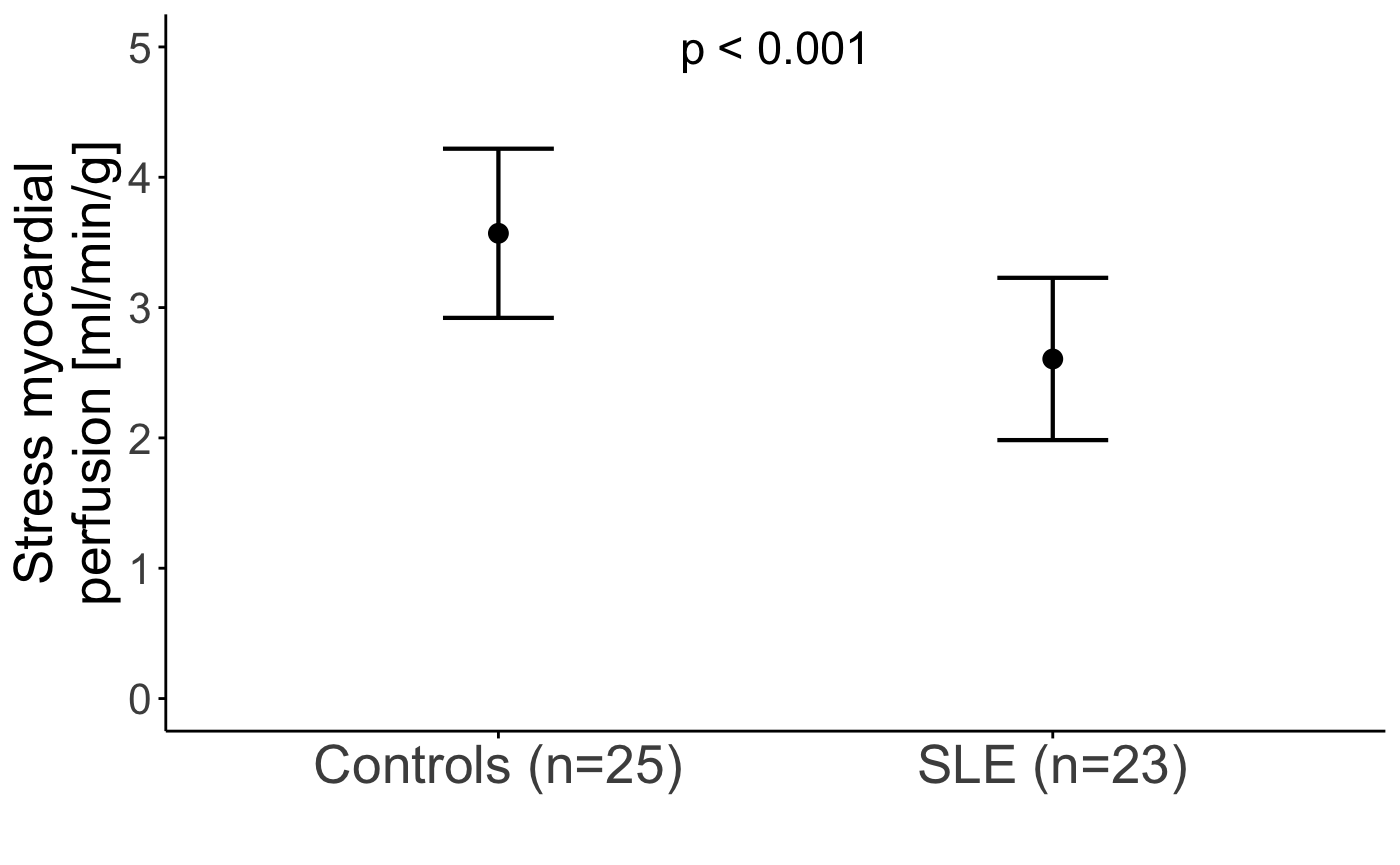
Figure 1. Two representative cases from pulmonary hypertension patients showing from left to right columns (a, e) combined gradient echo magnitude from NAV QSM, (b, f) combined magnitude from spiral QSM, (c, g) NAV QSM and (d, h) spiral QSM. The estimated differential blood oxygen saturation between the right/left heart (ΔSaO2) in subject #1 (first row) is 34% from NAV QSM and 33% from spiral QSM versus 36% from right heart catheterization (RHC). ΔSaO2 estimation in subject #2 (second row) is 28% from NAV QSM and 33% from spiral QSM versus 28% from RHC. 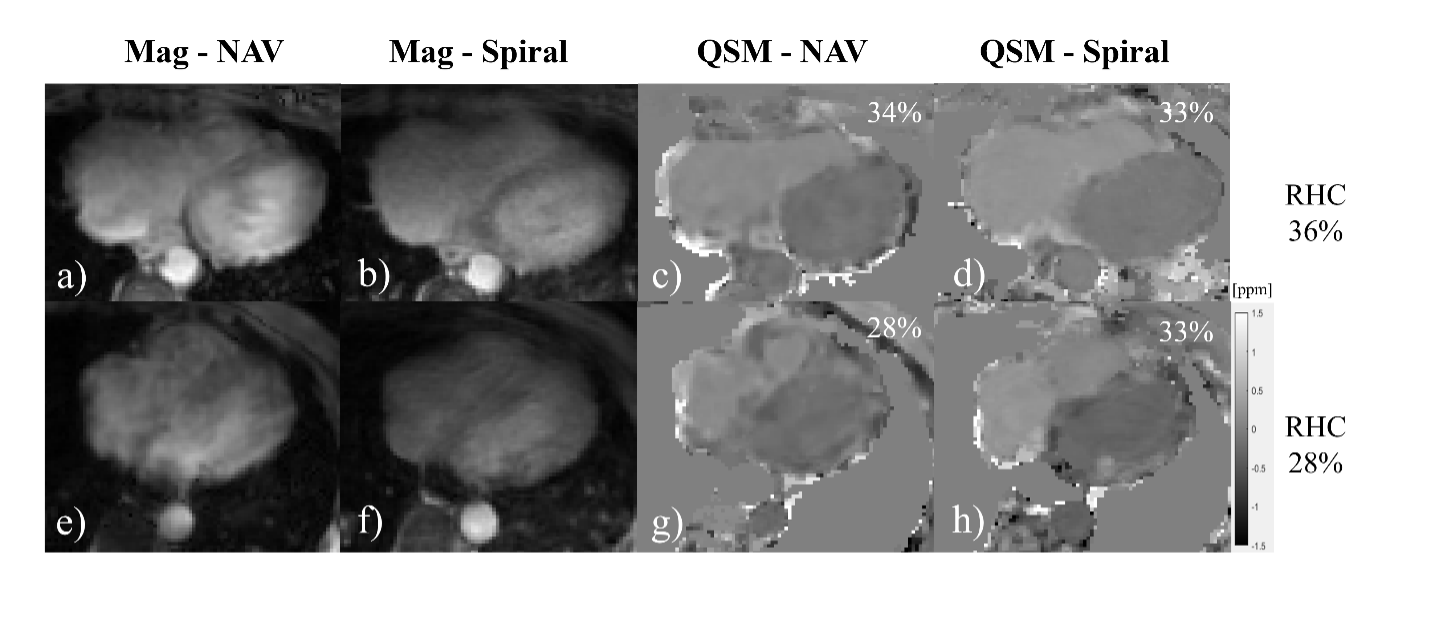
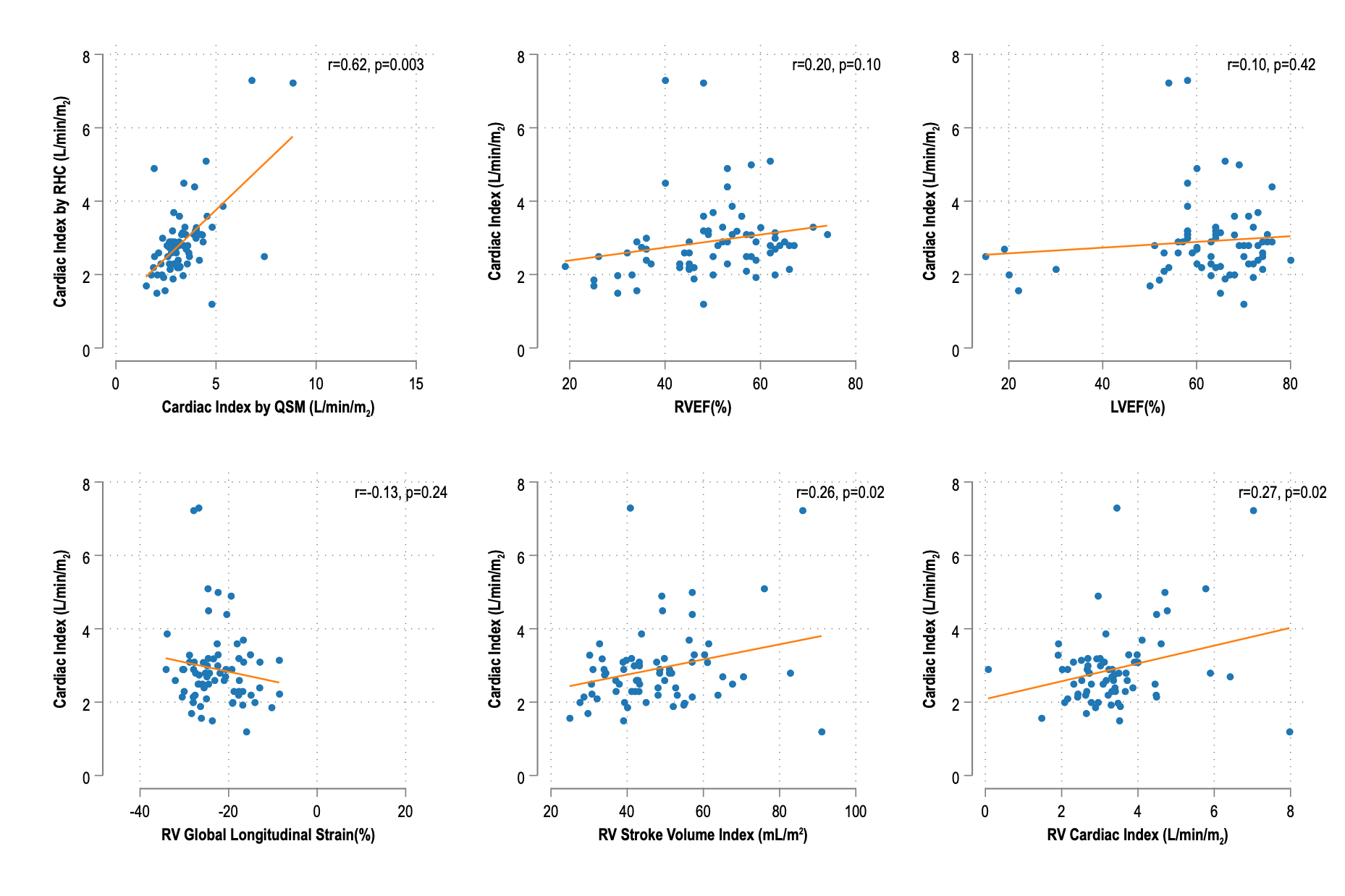
Scatter plots showing the relationship between cardiac index (L/min/m²) and various non-invasive cardiac magnetic resonance parameters.