Technologist Track
Joni Blood, RT
Cardiac MRI Technologist
Rady Children's Hospital
Joni Blood, RT
Cardiac MRI Technologist
Rady Children's Hospital
Ana E. Rodriguez Soto, PhD
Staff Scientist, Dept of Bioengineering
University of California San Diego

Francisco J. Contijoch, PhD, FSCMR
Associate Professor
University of California San Diego
Robyn Augustyn, BSc, RT
Advanced Imaging Technologist
Rady Children's Hospital
Daniel Vinocur, MD
Neuroradiologist
Rady Children's Hospital
Beth Feller Printz, MD
Pediatric Cardiologist
Rady Children's Hospital

Eleanor Lehnert Schuchardt, MD
Assistant Professor Health Sciences, Pediatric & Fetal Cardiology
University of California San Diego / Rady Children's Hospital
San Diego
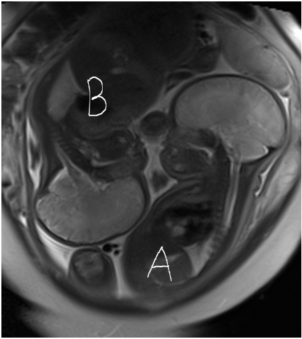
MRI was performed at 36 weeks gestation with patient supine on a 1.5-T (Sola, Siemens Healthineers, Erlangen, Germany) with the 18-channel body array and the inherent body/spine coils. Haste localizers in true planes were used to identify each fetus and define position. (Figure 1) Then the external cardiac Doppler gating device (NorthH Medical, Hamburg, Germany) was positioned on Baby A, on maternal right and breech.
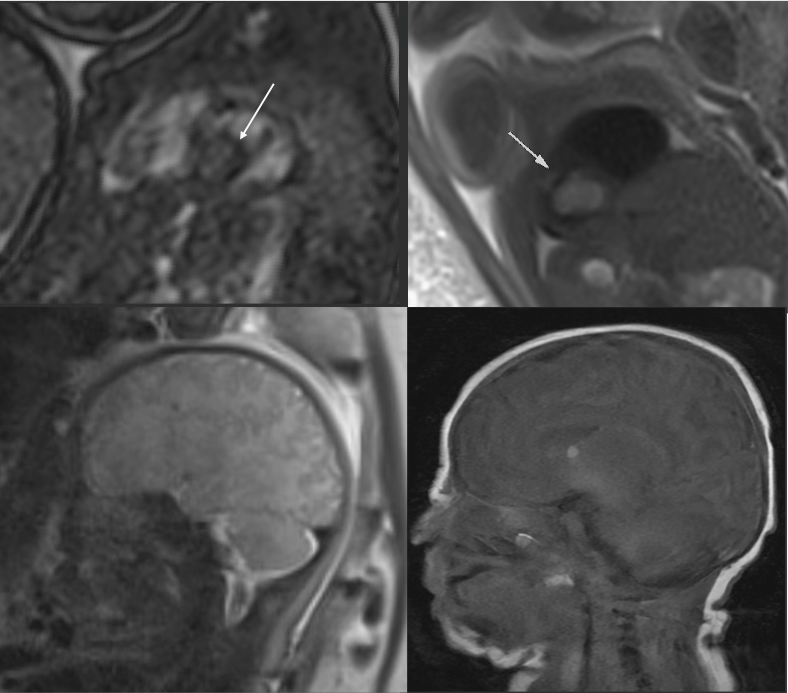
Brain imaging included T2-Haste in all three planes and DWI with ADC maps. Baby A had subtle T2 hypointense foci in the bifrontal white matter, hyperintense foci in the right ventricle and left foramen of Monro. Baby B had no intracranial abnormalities.
Cardiac imaging was directed only at Baby A and showed a large mass in the interventricular septum, which was isointense on T1 and hyperintense on T2. (Figure 2) Left ventricular function was mildly impaired as assessed by SAX Cine. Ejection fraction was 34%.
Overall findings were consistent with TS and aided definitive counseling of the expectant parents. Postnatal observation in the NICU was planned based on multisystem findings as well as plans for postnatal follow up with cardiology.
Baby A was confirmed to have a large cardiac rhabdomyoma in the interventricular septum, combined with central nervous system findings consistent with TS. Fetal MRI assisted with postnatal care planning by providing a more definitive design for what the child’s future medical needs would be. Baby A started on sirolimus postnatally, the larger ventricular rhabdomyoma has become significantly smaller, not causing any obstruction, and the baby has had no neurologic/developmental issues. The child has been followed by cardiology from birth.
Imaging both hearts and brains of twins at 36 weeks initially was a daunting task. We found it was feasible to image two CNS and one heart in approximately 90 minutes. The scan contributed to prenatal counseling.