Oral Case
SUMAN SINGHAL, MD
DIRECTOR CARDIAC IMAGING, HEAD OF DEPARTMENT
MAHAJAN IMAGING ,FORTIS ESCORTS HOSPITAL JAIPUR, India
SUMAN SINGHAL, MD
DIRECTOR CARDIAC IMAGING, HEAD OF DEPARTMENT
MAHAJAN IMAGING ,FORTIS ESCORTS HOSPITAL JAIPUR, India
RAHUL SINGHAL, MD
ADDITIONAL DIRECTOR CARDIOLOGY AND CARDIAC ELECTROPHYSIOLOGY
FORTIS ESCORTS HOSPITAL JAIPUR, India
79-year-old male, hypertensive, diabetic presented with history of dizziness associated with vomiting. He had similar complaints along with blurring of vision few days back. X-ray chest revealed raised right dome of diaphragm with old fibro-atelectatic bands in left upper lobe (old history of Koch's). ECG revealed normal sinus rhythm. Echocardiography revealed echogenic mass on posterior mitral leaflet (PML) protruding in left atrium causing restricted movements of mitral valve and mild mitral stenosis? thrombus? vegetation? myxoma.
On Cardiac MRI, for further assessment of cardiac mass, well defined, focal mass lesion along the posterior mitral annulus attached to PML. Mass was hypointense on T1W, T2W, T2FS images with no contrast enhancement on first pass gadolinium enhanced scan. Mild peripheral enhancement was seen on late gadolinium enhanced (LGE)scan. On concurrent CT images high attenuation calcified mass lesion was seen in posterior mitral valve with mild calcification along anterior mitral leaflet. Based on imaging findings of CT and CMR, diagnosis of caseous calcification of mitral annulus (CCMA) was given and patient was referred for surgery. On surgery ,encapsulated caseous calcification with necrotic centre of PML was found extending into posterior mitral annulus.Patient underwent debridement and excision of mass with mitral valve replacement .
Diagnostic Techniques and Their Most Important Findings:
Echocardiography revealed echogenic mass on posterior mitral leaflet (PML) measuring 2.3 cm x 1.8 cm protruding in left atrium causing restricted movements of mitral valve and mild mitral stenosis? thrombus? vegetation? Myxoma (Figure1).
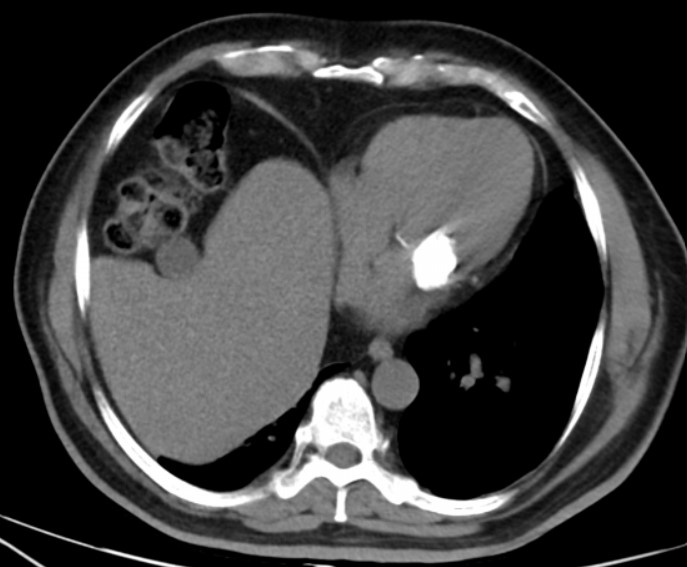
On Cardiac MRI, well defined, focal mass lesion along the posterior mitral annulus attached to PML measuring approximately 24 x 28 x 18 mm in size was seen. The mass was non-mobile more toward atrial side causing functional narrowing of mitral valve. Mass was hypointense on T1W, T2W, T2FS images with no contrast enhancement on first pass gadolinium enhanced scan. Mild peripheral enhancement was seen on late gadolinium enhanced (LGE) scan suggesting fibrous cap around avascular core of necrotic caseous material (Figure2). On concurrent CT images high attenuation calcified mass lesion was seen in posterior mitral valve with mild calcification along anterior mitral leaflet (Figure3). Based on imaging findings of CT and CMR, diagnosis of caseous calcification of mitral annulus (CCMA) was given. Close differentials are myxoma / fibroma which reveal intense enhancement on post-contrast scan.
Learning Points from this Case:
Caseous calcification of mitral annulus is rare form of degenerative mitral annular calcification (MAC) -0.062.07% of general population and typically involves the posterior mitral annulus and the atrioventricular groove. In early phase, mass is usually hyperintense on both T1 and T2W images due to high fluid content and liquefactive necrosis. Associated calcification within or around the mass appears hypointense on T1W and T2W images with no first pass contrast enhancement. Imaging feature of peripheral enhancement on LGE differentiates MAC from CCMA as it suggests fibrous cap around avascular core of necrotic caseous material. This led us to make definitive diagnosis of CCMA- proven on surgery. Indication of surgery is in larger lesions and in embolic events.
In summary, the primary imaging features of CCMA include posterior mitral valve annulus involvement, the presence of calcifications on CT, lack of first pass contrast enhancement on MRI with mild peripheral enhancement on LGE.
ECHOCARDIOGRAPHY SHOWING ECHOGENIC MASS ALONG POSTERIOR MITRAL LEAFLET CAUSING FUNCTIONAL NARROWING OF MITRAL VALVE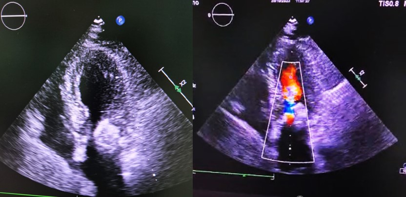
HYPOINTENSE MASS LESION ON T1W, T2W ( A,B) WITH PERIPHERAL ENHANCEMENT ON LGE SHORT AXIS (C) AND 4CHAMBER(D) IMAGES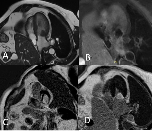
DENSE CALCIFIC HIGH ATTENUATION MASS ALONG POSTERIOR MITRAL VALVE