P/CHD
Thomas M. Vollbrecht, MD
Physician
University Hospital Bonn, Germany
Thomas M. Vollbrecht, MD
Physician
University Hospital Bonn, Germany
Luis F. Goncalves, MD
Section Head of Fetal Imaging
Phoenix Children's
Dianna Bardo, MD
Pediatric Radiologist
Lurie Children's Hospital of Chicago
Christopher Hart, MD
Pediatric Cardiologist
University Hospital Bonn, Germany
Alex J. Barker, PhD
Associate Professor
Children's Hospital Colorado, University of Colorado Anschutz Medical Campus

Richard M. Friesen, MD
Assistant Professor
Children's Hospital Colorado

Julian A. Luetkens, MD
Physician
University Hospital Bonn, Germany
Fetal cardiac cine MRI using Doppler US gating is an emerging imaging modality for assessing anatomy and function of the fetal heart. However, lack of evidence about parameters influencing image quality hinders full utilization of its potential. This study aimed to identify factors influencing image quality in a multi-center cohort of fetal cardiac MRI examinations.
Methods:
This multi-center study included fetal cardiac MRI scans between April 2021 and July 2023 at three different centers (University Hospital Bonn, Children’s Hospital Colorado, Phoenix Children’s Hospital). Based on a 5-point Likert scale (1=non-diagnostic to 5=excellent), image quality of axial cine stacks was assessed in three categories (contour sharpness, blood pool-to-structure contrast, level of artifacts). An overall image quality score was determined by the average of all criteria. Comparisons of image quality scores were performed between dichotomized groups for nine parameters with potential impact on image quality, including gestational age, body mass index (BMI), fetal motion, patient positioning, gating signal stability, breathing technique, field strength, slice thickness, and flip angle. Image quality scores between groups were compared using the Mann Whitney U test.
Results:
98 scans were analyzed (mean gestational age, 33.6 ± 4.5 weeks). Overall image quality was higher in subjects with: BMI < 30 kg/m2 compared to those with BMI ≥ 30 kg/m2 (4.4 ± 0.7 vs 4.1 ± 0.7, p = < 0.001); gestational age ≥ 32 weeks compared to those with gestational age < 32 weeks (4.3 ± 0.7 vs 4.1 ± 0.8, p = < 0.001); low severity of fetal motion compared to exams with severe fetal motion (4.5 ± 0.6 vs 3.9 ± 0.8, p = < 0.001); supine position of the mother compared to left lateral position (4.5 ± 0.5 vs 4.2 ± 0.8, p = 0.001); stable DUS gating signal compared to those with an unstable DUS gating signal (4.5 ± 0.6 vs 4.0 ± 0.8, p = < 0.001); flip angles > 60° compared to acquisitions with flip angles ≤60° (4.4 ± 0.6 vs 4.2 ± 0.8, p = 0.048). No difference in overall image quality was observed, but higher contour sharpness and fewer artifacts were present in scans using breath-holds compared to free breathing (5 [4-5] vs 4 [4-5], p = 0.042 and 4 [3-5] vs 4 [3-5], p = 0.014, respectively), whereas higher contrast and fewer artifacts were found at 3T compared to 1.5T field strength (5 [4-5] vs 5 [4-5], p = 0.041 and 4 [4-5] vs 4 [3-5], p = 0.010, respectively). No significant differences were found between cine stacks with 4 mm slice thickness compared to cine stacks > 4 mm (4.1 ± 0.9 vs 4.1 ± 0.9, p = 0.935).
Conclusion:
Various factors (e.g. BMI) influence image quality of fetal cardiac cine MRI with Doppler US gating. Understanding these factors might help achieving reliable examination results and better exploiting the potential of fetal cardiac MRI in clinical routine.
Stacked bar charts show the distribution of Likert scale scores used for overall image quality assessment. Likert scale scores for overall image quality were averaged between the individual ratings for the three categories contour sharpness, blood pool-to-structure contrast, and level of artifacts from all readers. P-values were calculated using the Wilcoxon.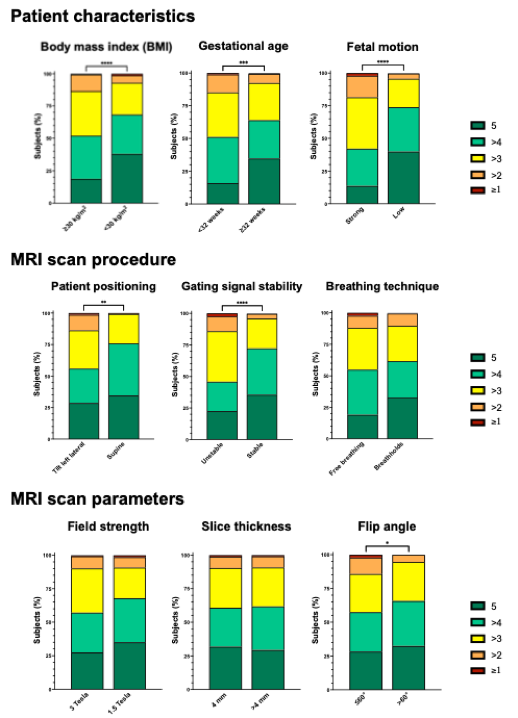
Balanced steady-state free precession cine image examples from the three centers University Hospital Bonn (Bonn), Children’s Hospital Colorado (Colorado) and Phoenix Children’s Hospital (Phoenix), each in one slice at ventricular level, outflow tract level, and aortic arch or ductus arteriosus level. The images from gestational weeks 36 and 24 (A) show the clear improvement of structure delineability as pregnancy progresses. Images of participants with different body mass index (B) demonstrate the influence of obesity on image quality resulting from poorer MR signal. Strong fetal movement (C) may render images non-diagnostic due to low contrast and pronounced artifacts, as seen here particularly in the slice at the outflow tract level.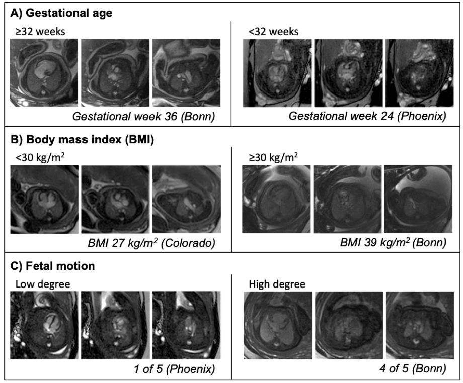
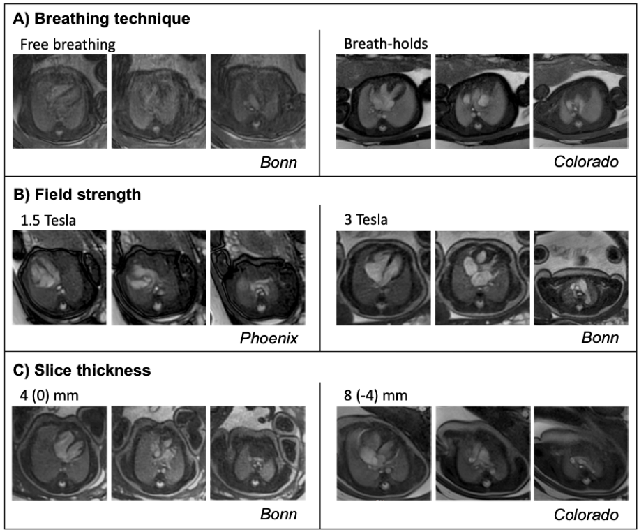
Balanced steady-state free precession cine image examples from the three centers University Hospital Bonn (Bonn), Children’s Hospital Colorado (Colorado) and Phoenix Children’s Hospital (Phoenix), each in one slice at ventricular level, outflow tract level, and aortic arch or ductus arteriosus level. Images acquired during free breathing (A) are inferior to those acquired during breath-holds because they are affected by poor contour sharpness and linear artifacts caused by maternal abdominal movements associated with breathing. Images acquired at 1.5T (B) had higher contrast and fewer artifacts; however, the overall image quality was not significantly different. When comparing the images by slice thickness (C), no differences were found in any of the three categories.