Clinical & Translation
Bastiaan J.C te Kiefte, MD
Drs.
Leiden University Medical Center, Netherlands
Bastiaan J.C te Kiefte, MD
Drs.
Leiden University Medical Center, Netherlands
Mitck Ramaekers, MD
Drs.
Maastricht University Medical Center, Netherlands
Bouke P. Adriaans, MD, PhD
Dr.
Maastricht University Medical Center, Netherlands

Joe F. Juffermans, MSc
Researcher PhD candidate
Leiden University Medical Center, Netherlands
Hans C. van Assen, PhD
Image analysis
Leiden University Medical Center, Netherlands
Arthur Scholte, MD, PhD
Dr.
Leiden University Medical Center, Netherlands
Joachim Wildberger, MD, PhD
Prof.
Maastricht University Medical Center, Netherlands
Simon Schalla, MD, PhD
Dr.
Maastricht University Medical Center, Netherlands

Hildo Lamb, MD, PhD
Radiology Professor
Leiden University Medical Center, Netherlands
.jpg "Jos J.M Westenberg, PhD photo")
Jos J.M Westenberg, PhD
Senior Researcher
Leiden University Medical Center, Netherlands
Elastin and collagen fibres define the elastic properties of the aorta. Alterations of these fibres during aging lead to mechanical changes, associated with aortic disease such as aneurysm and dissection. Regional variations in fiber orientation cause these regions to respond differently to hemodynamic effects. This study investigated the influence of hemodynamic parameters on structural and mechanical properties of the aorta in a healthy population, as assessed by CMR.
Methods:
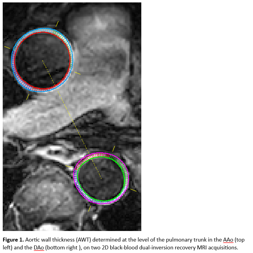
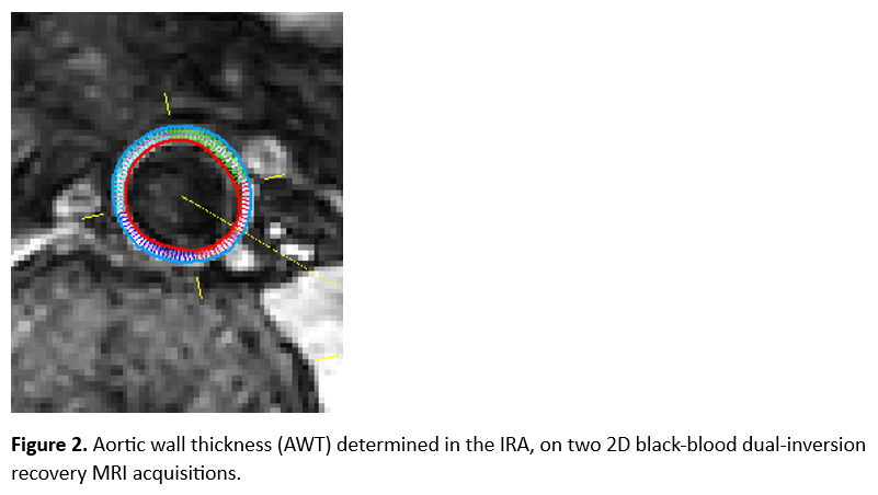
166 healthy volunteers (48% male) distributed in five age groups (1: 18-29, 2: 30-39, 3: 40-49, 4: 50-59, 5: 60-70 years) were included in this study and underwent CMR imaging of the entire aorta including 4D flow MRI sequences. The aorta was divided into three anatomical regions: ascending aorta [AAo], descending aorta [DAo], and infra-renal aorta [IRA]. Aortic wall thickness (AWT) was determined at the level of the pulmonary trunk in the AAo (AWTAAo) and the DAo (AWTDAo), and in the infra-renal aorta (AWTIRA), on two 2D black-blood dual-inversion recovery MRI acquisitions (Figure 1). PWV was assessed from 2D phase contrast MRI. Regional PWV was assessed by calculating a weighted average for all three regions. Flow displacement, peak systolic WSS (magnitude, axial, and circumferential), and WSS angle were also determined in all three regions.
Results:
PWVAAo, PWVDAo, and PWVIRA increase with age, with significant differences between age groups (P< 0.001), but not between sexes. Post-hoc analysis demonstrated that for age group 5 the PWV in all regions were significantly higher compared to all other groups (P< 0.001).
AWTAAo, AWTDAo, and AWTIRA increases with age, with significant differences between age groups (P< 0.001), but not between sexes. Post-hoc analysis demonstrated that for age group 5 the AWTAAo was significantly higher compared to age groups 1-3 (P< 0.001). For age group 5 the AWTDAo was significantly higher compared to all other groups (P< 0.001). AWTIRA was significantly higher for age group 5 compared to age groups 1 and 2 (P=0.001).
WSS angle is positively correlated with AWT in AAo for age groups 2 (r=0.42, P=0.009), 3 (r=0.40, P=0.015), and 5 (r=0.41, P=0.022). In DAo for age groups 1 (r=0.48, P=0.007), 3-5 (r=0.40, 0.49, 0.57, respectively) and IRA for age groups 3 (r=0.59, P< 0.001) and 5 (r=0.55, P=0.04). Maximum FD was only negatively correlated with AWTAAo for age group 2 (r=-0.39, P< 0.018). We did not find significant correlations between PWV and WSS angle, and between PWV and FD.
In multivariable analysis, WSS angle was the only parameter independently associated with increase in AWTAAo (ß=0.34, P< 0.001), in AWTDAo (ß=0.36, P< 0.001), and in AWTIRA (ß=0.29, P< 0.001).
Conclusion:
This study demonstrates that wall shear stress angle is associated with age related growth of aortic wall thickness, in a healthy population.