
Rapid Fire Abstracts
Leila Haririsanati, MD, MSc
Researcher
McGill University Health Center, Canada
Leila Haririsanati, MD, MSc
Researcher
McGill University Health Center, Canada
Moezedin javad Rafiee, MD
Research associate
Research Institute of the McGill University Health Center, Canada
Abhinav Sharma, MD
Assistant Professor
McGill, Canada

Matthias G. Friedrich, MD
Full Professor
McGill University Health Centre
Mc Gill University, Canada
Michael Chetrit, MD
Assistant professor
McGill University Health Center, Canada
.png)
Right bundle branch block (RBBB) causes a delay in the electrical conduction of the heart (1). It occurs in 0.2% to 1.3% of the population (2) and is considered a benign finding without any associated structural abnormalities (1).
Cardiovascular Magnetic Resonance (CMR) T1 Mapping has been recognized as a valuable tool for detecting changes in myocardial tissue characterization (3). Elevated native T1 values are associated with an increased water content (edema) or expansion of the interstitial space, e.g. in fibrosis (3, 4).
The impact of RBBB on myocardial tissue characterization in patients without previous history of cardiovascular disease has not been evaluated.
This study investigated the impact of RBBB on myocardial tissue characterization by native T1 mapping CMR imaging in participants without previous history of cardiovascular disease.
Methods:
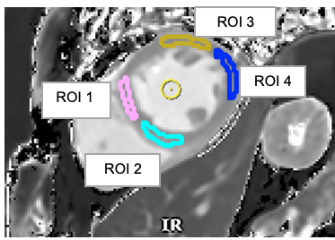
Twenty-one participants without a previous history of cardiovascular disease (43% female, mean age 58+/- 7.44 years old) with right bundle branch block in ECG, and eighteen control participants (50% female, mean age 56+/- 9.35 years old) were scanned on a 3T SIGNA Premier. Short axis T1 mapping with MOLLI-MOCO-DL T1 (Modified Look-Locker Inversion Recovery with motion correction and Deep Learning enhancement) sequence was applied. Four regions of interest (ROI) were drawn in a mid-ventricular slice (Figure 1) for each participant. T1 mapping values were measured using certified software.
Results:
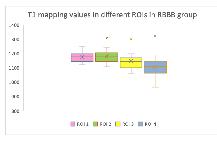
In the RBBB group, T1 values of the septum (ROI 1: 1175.48±50; ROI 2: 1181±53) were significantly higher than in the other regions (anterolateral/ROI 3: (1101.71±72) and inferolateral/ROI 4: 1106.47±76; p=0.0001 and 0.0001, respectively, Figure 2). There were no statistically significant differences between the two septal ROIs (p=0.85; Figure 2).
Septal T1 values in the RBBB group were also statistically higher than in the same myocardial region in the control group (ROI 1: 1144.39±31 ms, ROI 2: 1142.05±31 ms; p=0.0278 and 0.0107, respectively, Figure 3).
Conclusion:
In patients without a history of cardiovascular disease, Right Bundle Branch Block is associated with a septal increase of myocardial T1. The exact etiology of these changes needs further exploration.
Figure 1. Four regions of interest were drawn in mid-ventricular slice of left ventricle, representing anteroseptal (ROI 1, pink), infero-septal (ROI 2, turquoise), anterolateral (ROI 3, yellow), and inferolateral (ROI 4, blue) myocardium.
Figure 2: T1 values across septal (ROI 1 and 2) and anterolateral/inferolateral regions. The median septal T1 (ROI 1: 1175.48±50; ROI 2: 1181±53) was higher than that of the anterolateral (ROI 3: 1102±72) and the inferolateral (ROI 4: 1106±76) myocardium. There were no statistically significant differences between the T1 values of ROI 1 and ROI 2 (p=0.85), but both septal T1 values were significantly higher than anterolateral (ROI 3; p=0.026 and 0.0142) and inferolateral (p=0.0001 and 0.0001).
Figure3: T1 mapping values of the septum (ROI 1 and 2) in RBBB group were statistically higher than values of the control group (ROI 1: 1144.39±31 ms. ROI 2: 1142.05±31 ms) (p= 0.0278 and 0.0107). (*) denotes statistical significance.
