Early Career
Zi Wang, PhD
PhD
Xiamen University, China (People's Republic)
Zi Wang, PhD
PhD
Xiamen University, China (People's Republic)
Fanwen Wang
PhD student
Royal Brompton Hospital and National Heart and Lung Institute, Imperial College London, United Kingdom
Yan Li, MSc
Shanghai, China
Department of Radiology, Ruijin Hospital, Shanghai Jiao Tong University School of Medicine, China (People's Republic)

Chen Qin, PhD
Lecturer (Assistant Professor)
Imperial College London, United Kingdom
Jun Lyu, PhD
Boston, United States
Harvard Medical School
Ouyang Cheng, PhD
London, United King
Imperial College London, United Kingdom
Shuo Wang, PhD
Shanghai, China
Digital Medical Research Center, School of Basic Medical Sciences, Fudan University, China (People's Republic)
Yajing Zhang, PhD
Suzhou
Philips Healthcare, China (People's Republic)

Chengyan Wang, PhD
Associate Professor
Fudan University, China (People's Republic)
Chengyan Wang, PhD
Associate Professor
Fudan University, China (People's Republic)
Accelerated cardiac MRI is highly expected to achieve time-efficient and patient-friendly imaging, and then advanced image reconstruction approaches are required to recover high-quality, clinically interpretable images from undersampled measurements. However, the lack of publicly available cardiac MRI k-space dataset in terms of both quantity and diversity has severely hindered substantial technological progress. Here, we provide a standardized, diverse, and high-quality CMRxRecon2024 dataset to facilitate the technical development, fair evaluation, and clinical transfer of cardiac MRI reconstruction approaches, towards promoting the universal machine learning that enable fast and robust reconstructions across different cardiac MRI protocols in clinical practice (Figure 1).
Methods:
Figure 2 shows the overall workflow to prepare our CMRxRecon2024 dataset from data acquisition to the final released dataset.
The study received approval from our local institutional review board (approval number: MS-R23). As part of the written consent process, participants agreed to make their anonymized data publicly available. Between June 2023 and February 2024, 330 healthy volunteers (156 males and 174 females) provided written informed consent and participated in the study.
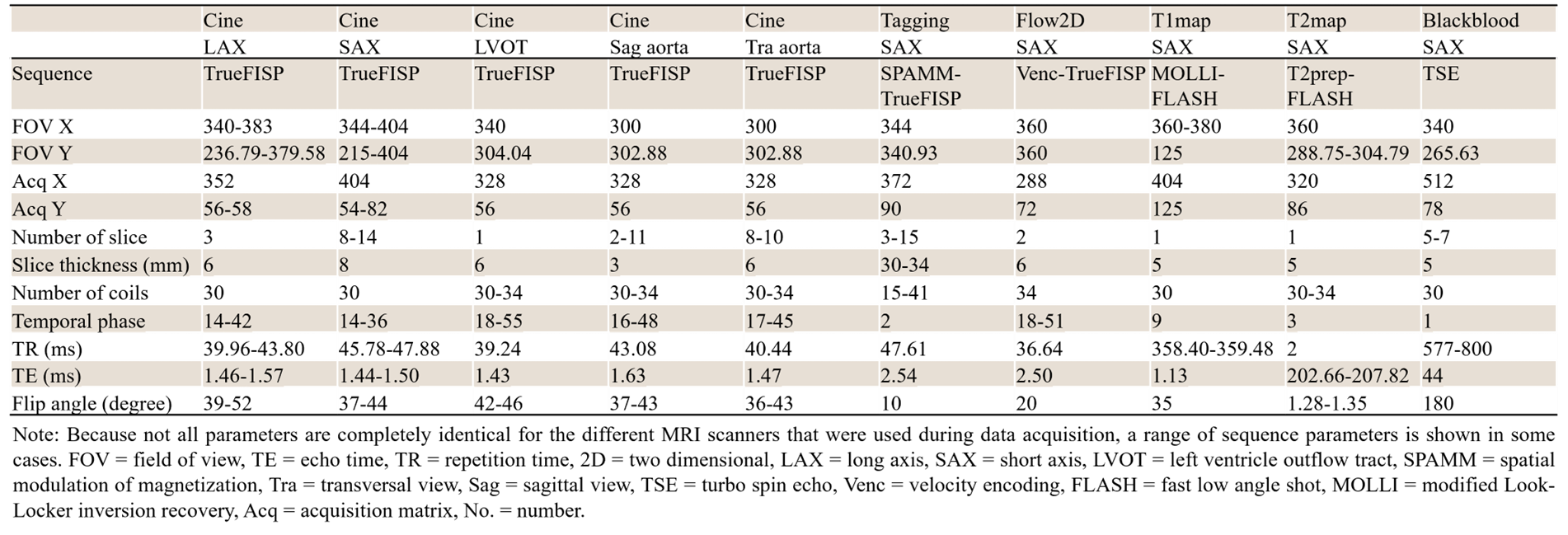
Data were acquired using a 3T scanner (MAGNETOM Vida, Siemens Healthineers), equipped with dedicated multi-channel cardiac coils. The typical acquisition parameters of imaging protocols are summarized in Figure 3.
To simulate different acceleration scenarios, various k-space undersampling trajectories (i.e., uniform, Gaussian, and pseudo radial) with different acceleration factors (i.e., 4~24) were provided for retrospective undersampling.
Results:
To date, the released CMRxRecon2024 dataset is the largest and most diverse publicly available cardiac k-space dataset. It is acquired from 330 healthy volunteers, covering commonly used modalities (cardiac cine, T1/T2 mapping, tagging, phase-contrast, and black-blood imaging), anatomical views (long-axis with 2-chamber, 3-chamber, and 4-chamber, short-axis, left ventricle outflow tract, and aorta with transversal and sagittal views), and acquisition trajectories (uniform, Gaussian, and pseudo radial sampling with different acceleration factors) in clinical cardiac MRI workflows. The CMRxRecon2024 dataset can be downloaded from the Synapse repository at https://www.synapse.org/#!Synapse:syn54951257/wiki/627141.
Moreover, to facilitate data usage, advanced method development, and fair performance evaluation, the tutorials, benchmarks, and data processing tools are provided in the Github repository: https://github.com/CmrxRecon/CMRxRecon2024.
Conclusion:
In summary, CMRxRecon2024 dataset is of significant benefit for accelerating the deployment of advanced machine learning methods and for the ultimately clinical adaption, to achieve more time-efficient, patient-friendly, and reliable diagnosis of cardiovascular diseases.
Figure 1: An overview of the released CMRxRecon2024 dataset. The dataset includes multi-modality cardiac MRI with diverse anatomical views. With various under sampling trajectories and acceleration factors, deep learning-based reconstruction methods can be developed for high spatiotemporal resolution images and comprehensive cardiac assessment in reduced scanning time.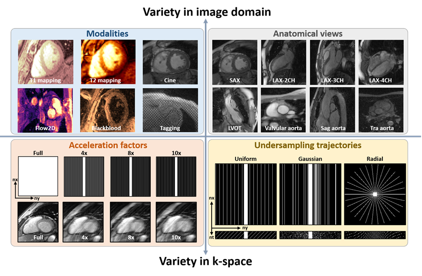
Figure 2: The workflow to prepare CMRxRecon2024 dataset from data acquisition to the final released dataset. Multi-coil, multi-modality, and multi-view k-space data were acquired from 330 healthy volunteers using a 3T MRI scanner with multi-channel cardiac coil.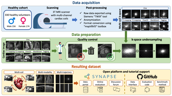
Figure 3: Acquisition parameters for the imaging protocols used to acquire k-space data represented in the CMRxRecon2024 dataset.