
Rapid Fire Abstracts
Bryan Gonzalez, BEng
R&D Devops Engineer
Children's National Hospital
Bryan Gonzalez, BEng
R&D Devops Engineer
Children's National Hospital
Ryan P. O'Hara, PhD
Postdoc
Children's National Hospital
Francesco Capuano, PhD
Assistant Professor
Universitat Politecnica deCatalunya Barcelona Tech, Spain
Aybala Tongut, MD
Cardiac Surgeon
Children's National Hospital
Menan Desai, MD
Cardiac Surgeon
Children's National Hospital

Yue-Hin Loke, MD
Associate Professor
Children's National Medical Center
Patients with congenital heart disease (CHD) undergo advanced cardiac imaging to plan for surgery. In addition to conventional visualization on a 2D screen, anatomical visual aids such as 3D models have been used for surgical planning. However, contemporary 3D modeling of complex CHD is limited by the lack of dynamic visualization of cardiac structures and cardiac flow. Novel CMR sequences such as 4D flow now allow for dynamic visualization of cardiac anatomy and flow. This study evaluates the potential of Virtual Reality (VR) to visualize 4D flow as an alternative, analyzing its impact on overall efficiency and quality of implementation against conventional 2D and 3D methods.
Methods:
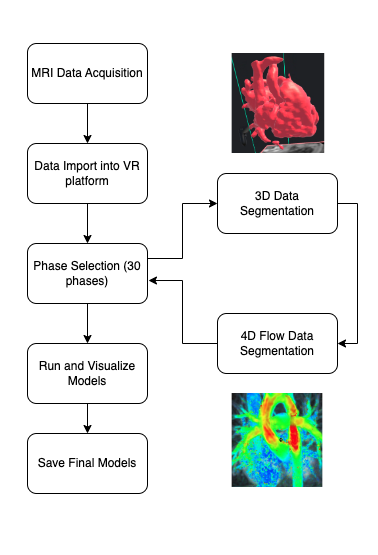
Anonymized patient cases with different types of CHD were retrospectively analyzed. Each patient had one ferumoxytol-enhanced 4D flow dataset that was loaded for segmentation. The case was segmented with using conventional workstation-based software (Slicer 3D, Mimics) or VR-based segmentation techniques (Elucis, Realize Inc). For the conventional-based method, one static model was created from a single diastolic phase within the 4D flow dataset. For VR-based segmentation, the dynamic 3D model across all cardiac phases within the 4D flow dataset was segmented. Additionally, relevant flow fields within phase-contrast data was also highlighted and segmented (Fig 1). Two primary outcomes were time to complete segmentation and diagnostic quality of the resultant 3D model as determined by physician.
Results:
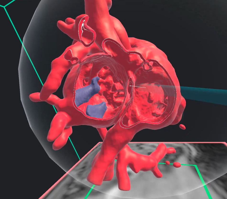
Twenty individual cases were anonymized and analyzed (Table 1). The conventional-based method was performed over an average of 71 ± 23 minutes. Using the VR-based methodology platform, each case took an average of 73 ± 15 minutes to be completely segmented. The conventional-based method was able to demonstrate the relevant cardiac anatomy, whereas the VR-based method also demonstrated relevant pathologic flow features (e.g. atrioventricular valve regurgitation, Fig 2).
Conclusion:
Visualization of 4D flow can be enhanced using a Virtual Reality based segmentation software. The efficacy of this methodology will continue to be investigated with direct feedback from the cardiac surgeons from this institution.
Figure 1 Caption - Workflow outlining the process of capturing MRI data, segmenting both 3D and 4D datasets for each phase, and visualizing 4D flow in a Virtual Reality environment.
Figure 2 Caption – Screenshot showing 4D flow visualization within a 3D heart model using a virtual reality platform. Atrioventricular valve regurgitation is shown as the blue structure inside the heart.
