
Quick Fire Cases
Calder Sheagren, BSc
Graduate Student
University of Toronto, Canada
Calder Sheagren, BSc
Graduate Student
University of Toronto, Canada
Terenz Escartin, MSc, BSc, MRT
Sunnybrook Research Institute & University of Toronto, Canada
Nasim Shadafny, MD
Fellow
Sunnybrook Health Sciences Centre, Canada
Maria Terricabras Casas, MD
Electrophysiologist
Sunnybrook Health Sciences Centre, Canada
Stephanie Poon, MD, MSc
Associate Professor
Sunnybrook Health Sciences Centre, Canada
Idan Roifman, MD, MSc, FSCMR
Assistant Professor
University of Toronto, Canada

Graham Wright, PhD
Professor and Sr. Scientist
Sunnybrook Research Institute and University of Toronto, Canada
Christopher Cheung, MD, MPH
Electrophysiologist
Sunnybrook Health Sciences Centre, Canada
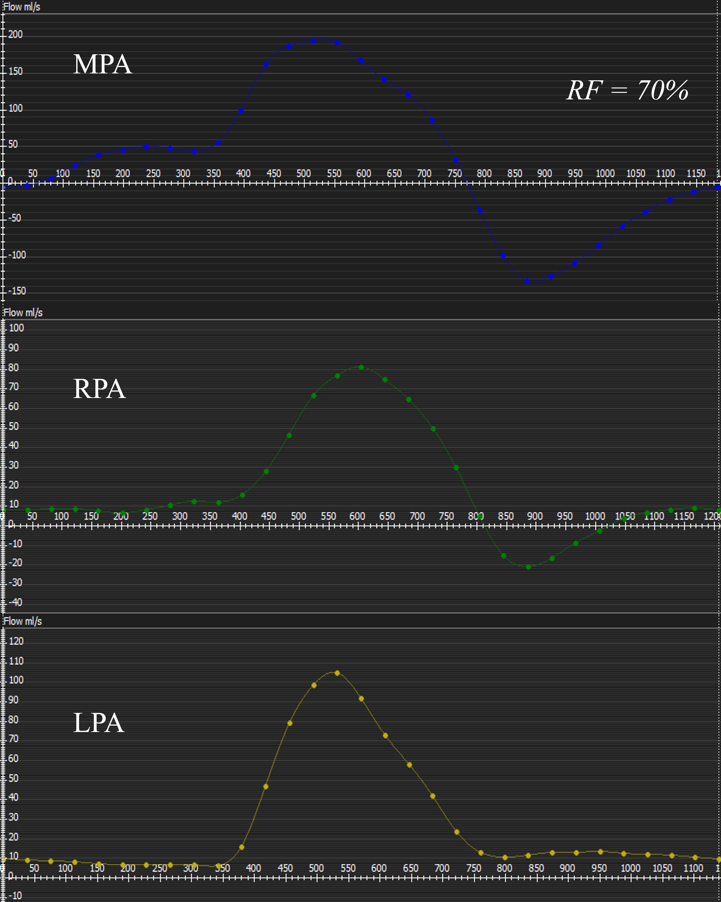
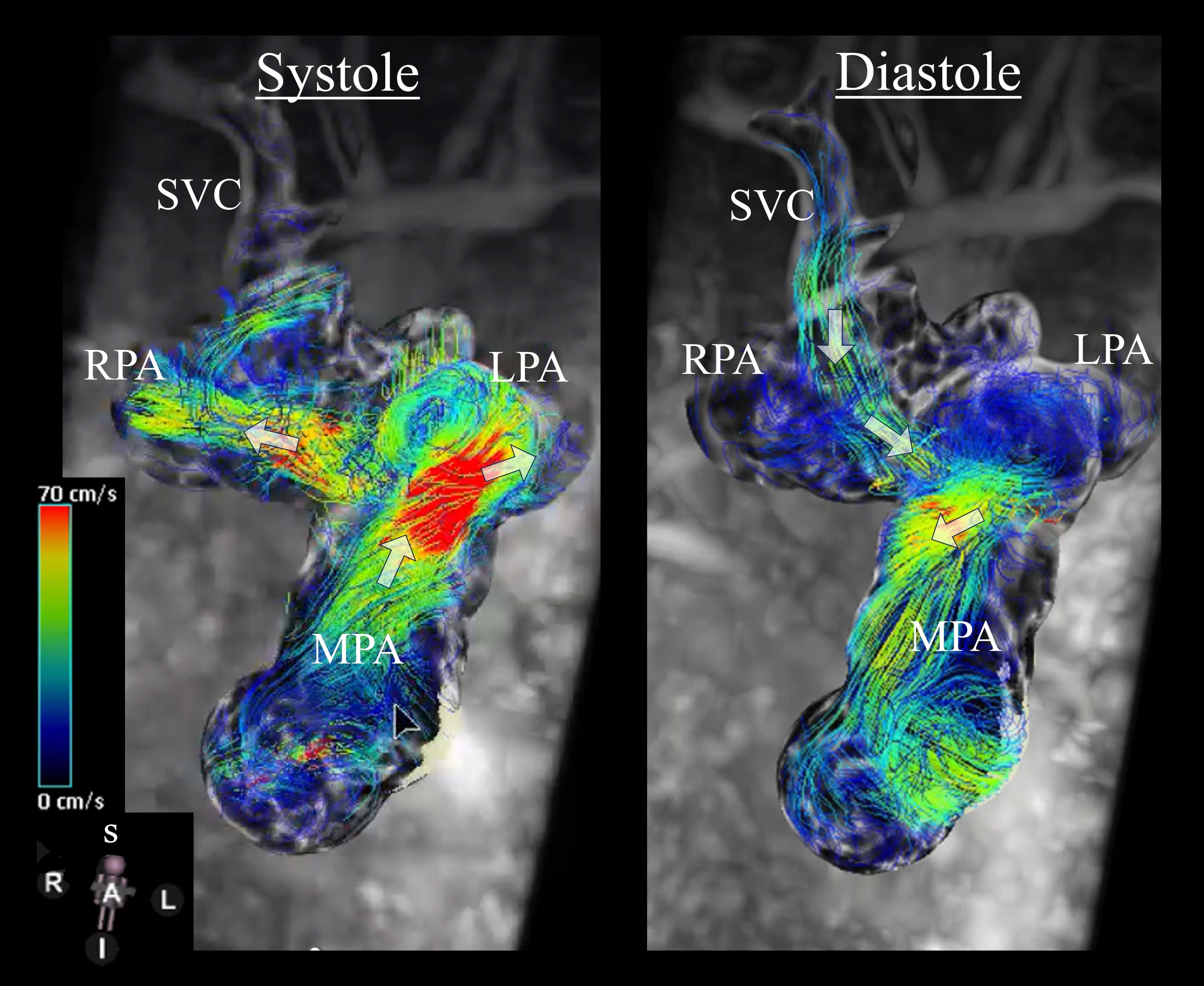
Echocardiography demonstrated severe pulmonary regurgitation, mild right ventricular (RV) dilation and normal RV and LV function. A CMR was performed to quantify the degree of pulmonary regurgitation and RV dilation. This demonstrated severe pulmonary regurgitation with a 70% regurgitant fraction on 2D and 4D phase contrast (PC) MRI yet no holodiastolic flow reversal in the branch pulmonary arteries. Flow pattern visualization on 4D PC demonstrated that BDG flow accounted for the majority of the pulmonary regurgitation and that the antegrade flow across the main pulmonary artery comprised half of the systemic venous return and the other half was supplied by the Glenn anastomosis. There was only mild RV dilation (indexed end-diastolic volume 102.4 ml/m2) and normal systolic function with ejection fraction of 56%. The BDG and the branch pulmonary arteries were unobstructed.
Learning Points from this Case: 1.5 ventricle circulation is an alternative to the Fontan procedure in the setting of a hypoplastic subpulmonic ventricle. The inferior vena cava flow is actively pumped by the RV and the lungs receive passive flow from the superior vena cava (SVC). This may result in an inefficient circulation with competing sources of pulmonary blood flow. 4D PC MRI allowed for comprehensive examination of blood flow patterns in this circulation. It quantified the degree of pulmonary regurgitation and explained the mechanism by showing the contribution of pulsatile and passive circulation to pulmonary blood flow. The mild degree of RV enlargement despite substantial pulmonary regurgitation was posited due to RV diastolic dysfunction and small RV size associated with PA/IVS. The primary cardiologist discussed pulmonary valve replacement with the patient and family through a shared decision-making model, and the case was subsequently discussed at a multidisciplinary surgical conference, where there was consensus to continue close observation due to the lack of symptoms, normal exercise capacity, and strong family preference to avoid intervention. She remains clinically well with close observation of symptoms, RV size, and RV function.
Figure 1. 2D phase contrast flow profiles in the main pulmonary artery(MPA, top), right pulmonary artery (RPA, middle) and left pulmonary artery (LPA, bottom) demonstrate severe pulmonary regurgitation with a 70% regurgitant fraction (RF), but only brief flow reversal in the branch pulmonary arteries.
Figure 2. Cardiac magnetic resonance imaging 4D flow stream lines in systole (A) and diastole (B) demonstrate superior vena cava flow as the predominant source of pulmonary regurgitant flow. Flow directionality is indicated by white arrows.
